Genitourinary Interventions
1. Percutaneous nephrostomy
A percutaneous nephrostomy tube is a small catheter that is placed through the skin into the kidney to drain urine that is blocked at some point in the urinary system. Urine flows into a bag outside the body. A blockage in the ureter or bladder is usually caused by a stone, scarring from infection or surgery, or cancer. Such obstruction is serious and may lead to kidney damage or severe infection. A percutaneous nephrostomy is performed with the patient lying on his or her stomach. An ultrasound (US) probe and real-time x-rays (fluoroscopy) are used to locate the kidney through the back and guide the placement of a needle through the skin into the kidney urinary system after application of a local anesthetic. Then, a small tube is placed allowing the obstructed urine to drain freely outside the body. A nephrostomy tube may be temporary or permanent depending on the indication and related medical condition.
Sedation: Local anesthesia (lidocaine) and moderate sedation (fentanyl and midazolam).
Procedure time: 30 minutes.
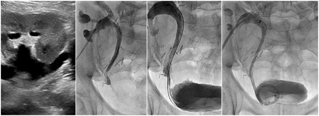
2. Nephroureteral and ureteral stent placement
A nephroureteral stent is a small tube that extends from outside the body into the kidney, down the ureter, and into the bladder. The function of this tube is to allow urine produced by the kidney to either drain freely into the bladder or outside the body into a bag preventing the buildup of urine within the kidney. This procedure is performed in patients who have developed a blockage in the ureter, the tube in the body that carries urine from the kidneys to the bladder. A blockage in the ureter is usually caused by a stone, scarring from infection or surgery, or cancer. The buildup of urine may damage the kidney and/or lead to serious infection. The procedure is performed with the patient lying on their stomach. An ultrasound (US) probe and real-time x-rays (fluoroscopy) are used to locate the kidney through the back and guide the placement of a needle through the skin into the kidney urinary system after application of a local anesthetic and sedation. Then, a small wire is guided from the kidney, down the ureter, and into the bladder over which the nephroureteral stent is placed, thus opening the obstructed ureter and allowing urine to drain freely. Ultimately, the nephroureteral stent may be removed, exchanged for a stent that remains completely inside the body (ureteral stent), or left in place permanently.
Sedation: Local anesthesia (lidocaine) and/or moderate sedation (fentanyl and midazolam).
Procedure time: 30 minutes.