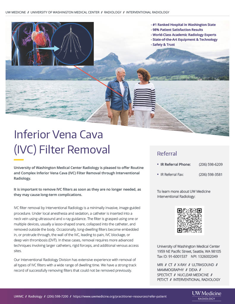
Inferior Vena Cava (IVC) Filters
1. Inferior vena cava filter placement
The deep veins of the body carry blood to the heart to be oxygenated by the lungs. Deep vein thrombosis (DVT) occurs when a thrombus (blood clot) forms in one or multiple deep veins in the body, typically in the legs. DVT blocks the blood flow and causes pain and swelling in the affected limb. DVT is a serious condition as thrombus has the potential to break off and travel to the lungs (pulmonary embolism), which may be life-threatening. The standard treatment for DVT includes blood thinning medications (anticoagulation) which prevents further clot formation or embolism. Blood thinners, however, do not work in all patients, and some patients cannot take them due to a pre-existing health condition. In these instances, placement of an inferior vena cava (IVC) filter may be indicated. An IVC filter is a small, umbrella-like device that is placed within the inferior vena cava (central vein in the abdomen) that can trap blood clots before they reach the lungs. An Interventional Radiologist may insert an IVC filter with minimally invasive techniques by accessing a vein in the neck or groin and position the IVC filter in the vein using real-time x-rays (fluoroscopy). The IVC filter may be left in permanently or removed when it is no longer needed. It is important to discuss the long-term plan for the IVC filter with a doctor (see IVC filter removal below).
Sedation: Local anesthesia (lidocaine) and moderate sedation (fentanyl and midazolam).
Procedure time: 30 minutes.
2. Routine inferior vena cava filter removal
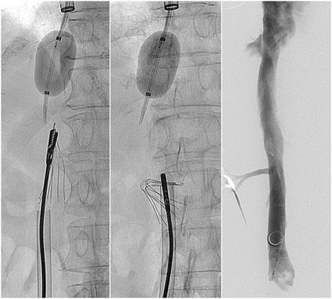
For most patients, it is important to remove inferior vena cava (IVC) filters as soon as they areno longer needed, as these devices may cause long-term complications. Undergoing removal of an IVC filter is similar to the initial placement. Filter removal is a non-surgical, minimally invasive procedure. Radiographic (x-ray) images will be obtained of the IVC filter to determine its exact location. The Interventional Radiologist will then use one or multiple devices, usually a snare (lasso), to grasp the tip of the filter, collapse the filter and remove it through a catheter. Additional images are then obtained to confirm the filter was removed in its entirety.
Sedation: Local anesthesia (lidocaine) and moderate sedation (fentanyl and midazolam).
Procedure time: 30-60 minutes.
3. Complex inferior vena cava filter removal
Occasionally, IVC filters become fixed within the wall of the IVC or the filter breaks into multiple pieces. In these cases, removal requires more advanced minimally invasive techniques and is best performed at centers with highly experienced specialists. Computed tomography (CT) images may be obtained for pre-procedure planning. The filter is typically removed through a larger catheter inserted into the neck, groin, or both. Using a variety of tools including snares (lasso), small forceps, or a laser, the filter may be carefully removed through a catheter under real-time radiographic (x-ray) guidance.
Sedation: Local anesthesia (lidocaine) and moderate sedation (fentanyl and midazolam) or general anesthesia while completely asleep with an endotracheal tube.
Procedure time: 30-120 minutes.