Venous Access
1. Peripherally inserted central venous catheter
A peripherally inserted central venous catheter (PICC) is a thin, flexible tube that is inserted by an Interventional Radiologist into a vein in the upper arm (or leg) and threaded into a large central vein near the heart. A PICC provides a safe route for intravenous medications, parenteral nutrition, or blood draws and avoids the need for repeated needle sticks. Compared to a standard intravenous (IV) catheter, PICC lines are ideal for treatment lasting several weeks or longer because they are more durable and comfortable. This procedure may be performed in a procedure suite or at the bedside. A PICC may be used for 2-6 weeks before removal.
Sedation: Local anesthesia (lidocaine) and/or moderate sedation (fentanyl and midazolam).
Procedure time: 15 minutes.
2. Non-tunneled central venous catheter
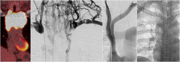
A central venous catheter (CVC), also called a “central line”, is a long, flexible tube that is inserted by an Interventional Radiologist through one of the large veins found in the neck, upper chest, or groin to allow access to the central bloodstream. A CVC is longer than a standard intravenous catheter (IV) and can remain in the body for a longer time. Unlike a tunneled device, a non-tunneled CVC is appropriate for short treatment periods (days to several weeks). In most circumstances, a non-tunneled CVC can be placed in the intensive care unit, and a chest x-ray is taken to confirm its position. When performed by interventional radiologists, ultrasound and x-ray (fluoroscopy) images are used to guide the procedure.
Sedation: Local anesthesia (lidocaine) and/or moderate sedation (fentanyl and midazolam).
Procedure time: 30 minutes.
3. Tunneled central venous catheter
Similar to non-tunneled CVCs, a tunneled CVC is a long, flexible tube which is placed by an Interventional Radiologist into a large vein in the neck, upper chest, or groin and situated under the skin to allow access to the central bloodstream. Tunneling the catheter under the skin protects the catheter from becoming infected and allows better fixation of the catheter to the tissues, so it may be used for longer periods of time (e.g. months) compared to non-tunneled CVCs. Ultrasound and x-ray images are used to guide the procedure.
Sedation: Local anesthesia (lidocaine) and/or moderate sedation (fentanyl and midazolam).
Procedure time: 30 minutes.
4. Port-a-Cath or Port placement
Similar to CVCs, a port is a special reservoir that is attached to a flexible tube which is placed by an Interventional Radiologist into a large vein in the neck. The port is placed under the skin to prevent it from becoming infected. Ports are best suited for infusions requiring intermittent access over a long periods of time (e.g. months) compared to daily infusions or short-term use.
Sedation: Local anesthesia (lidocaine) and/or moderate sedation (fentanyl and midazolam).
Procedure time: 50 minutes.