Venous Samplings
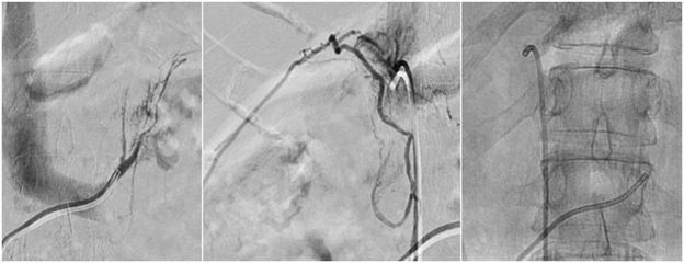
1. Adrenal venous sampling
High blood pressure (hypertension) is a common medical condition that is often well controlled with medications. Rarely, multiple medications are ineffective indicating that there may be another underlying cause of high blood pressure. In a small percentage of patients, high blood pressure is caused by release of too much aldosterone, a hormone produced by the adrenal glands, located above the kidneys. This condition is known as a condition called primary aldosteronism. Most often, that disorder is caused by either overproduction of the hormone by both glands or release of excess hormone by a benign tumor on one gland (Conn syndrome). The former is treated with drug therapy, but the latter is best treated by removal of the gland. To make the distinction, blood from the adrenal veins can be sampled individually. To undergo adrenal vein sampling, a small catheter is inserted into a vein in the groin or neck after administering local anesthesia (lidocaine). The catheter is then placed within the left and right adrenal veins, and blood samples are taken. This blood is then analyzed to determine the level of aldosterone hormone and identify the abnormal adrenal gland(s).
Sedation: Local anesthesia (lidocaine) and moderate sedation (fentanyl and midazolam).
Procedure time: 60-120 minutes.
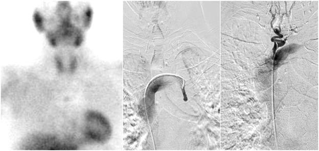
2. Parathyroid venous sampling
A parathyroid adenoma is a benign tumor of the parathyroid glands which are located in the back of the thyroid gland in the neck. Parathyroid adenomas produce an excess amount of parathyroid hormone (PTH) which may cause hyperparathyroidism. Surgical resection of the parathyroid adenoma is successful in relieving symptoms of this disorder. In rare cases, the parathyroid adenoma cannot be located during surgery or with standard imaging studies. In these rare cases, sampling of the parathyroid veins may be performed to locate the parathyroid adenoma. The procedure involves placing a small catheter into the various veins of the neck and chest to sample the blood and determine the location of the parathyroid adenoma. This information can then guide future surgery to remove the hormone-secreting tumor.
Sedation: Local anesthesia (lidocaine) and moderate sedation (fentanyl and midazolam).
Procedure time: 60-120 minutes.
3. Petrosal venous sampling
Cushing’s syndrome results from excessive production of the hormone cortisol. Cortisol is normally produced within the adrenal gland in the abdomen. A tumor within the pituitary gland, however, may cause the adrenal gland to produce excess cortisol. This procedure is used to determine if the source of the excessive hormone is the pituitary gland or somewhere else, often the lungs or abdomen. A catheter is inserted through the groin and navigated under x-rays (fluoroscopy) to the veins that drain the pituitary gland in the head (petrosal sinuses). Once the catheters are in place, blood samples are taken to determine the source of the excessive hormone. This information can then guide future surgery to remove the hormone-secreting tumor.
Sedation: Local anesthesia (lidocaine) and moderate sedation (fentanyl and midazolam).
Procedure time: 60-120 minutes.
4. Ovarian venous sampling
Hyperandrogenism is a medical condition that is characterized by high blood levels of androgens (male sex hormones such as testosterone) in females, most commonly caused by polycystic ovarian syndrome (PCOS). In rare cases, hyperandrogenism is caused by a tumor that secretes androgens. These tumors are typically identified on medical imaging including magnetic resonance imaging (MRI), computed tomography (CT), or ultrasound (US). When it is challenging to visualize the tumor on these scans, ovarian venous sampling may help diagnose and locate an androgen secreting tumor. In this procedure, a catheter is positioned within the various veins of the pelvis, and blood is sampled and tested for androgens.
Sedation: Local anesthesia (lidocaine) and moderate sedation (fentanyl and midazolam).
Procedure time: 60-120 minutes.
5. Hepatic venous sampling
Insulinoma is a rare tumor that secretes insulin. Similarly, nesidioblastosis is an abnormal increase of cells with abnormal increased insulin production that can form after gastric bypass surgery. Most often these conditions are benign; however, the high levels of insulin can cause life-threatening low blood sugar. Therefore, insulinomas should be surgically removed. Typically, these tumors are small (< 2 cm) and reside in the pancreas. Locating these tumors on imaging studies may be challenging, and sometimes a more invasive test called selective intra-arterial calcium stimulation with hepatic venous sampling is required to determine the tumor location. In this procedure, one catheter is positioned within a hepatic vein for blood sample collection, while another catheter is advanced to various locations along the mesenteric and hepatic arteries to inject calcium to stimulate insulin production from any insulinoma supplied by that artery. This information can guide future therapy including surgery to remove the insulin-producing tumor.
Sedation: Local anesthesia (lidocaine) and moderate sedation (fentanyl and midazolam).
Procedure time: 60-120 minutes.