Lymphatic Interventions
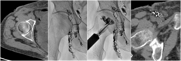
1. Lymphangiography
The lymphatic system is a network of tiny vessels in the body that drains clear or milky tissue fluid called lymph. The lymphatic system also consists of glands and organs including the lymph nodes, spleen, liver, and intestines. Lymphangiography is a minimally invasive, non-surgical technique that allows precise imaging of the lymphatic system and lymph nodes, usually to detect cancer spread or leakage of lymphatic fluid somewhere in the body. This procedure is done after application of a local anesthetic (lidocaine). The procedure is performed by an Interventional Radiologist using ultrasound (US) to guide a needle into a lymph node in the groin. Oily contrast material is injected into the lymph node which disperses and highlights the lymphatic vessels throughout the body. Images are taken using read-time x-rays (fluoroscopy) and are helpful for diagnostic and therapeutic purposes.
Sedation: Local anesthesia (lidocaine) and moderate sedation (fentanyl and midazolam) or general anesthesia (completely asleep with an endotracheal tube).
Procedure time: 60-180 minutes.
2. Lymphocele sclerosis and embolization
The lymphatic system is a network of vessels in the body that drains clear or milky tissue fluid called lymph. After undergoing surgery in the abdomen or pelvis, patients rarely develop a small collection filled with lymphatic fluid, called a lymphocele. Because lymphoceles can become infected or cause pain, most of them require direct treatment. A small catheter is placed through the skin into the collection so it can be drained outside the body into a bag. When the fluid has been drained, the cavity is filled with a sclerosing agent which will close the cavity and prevent it from returning. Complete treatment often requires periodic injections of sclerosing agent over weeks and rarely months.
Sedation: Local anesthesia (lidocaine) and/or moderate sedation (fentanyl and midazolam).
Procedure time: 30-60 minutes.
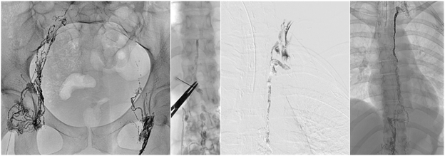
3. Thoracic duct disruption and embolization
The thoracic duct is a long tubular structure in the chest that carries clear or milky fluid called lymph from the chest and abdomen back into the venous system to be recirculated by the blood. Congenital abnormalities or surgical complications can result in a persistent leakage of lymph from the thoracic duct into the space around the lungs, producing a chylothorax. This condition may cause shortness of breath or malnutrition, as lymph is rich in proteins and fats. In these cases, a thoracic duct embolization procedure may be indicated. The site of leakage is identified and then closed off with embolization material such as tiny coils and glue. This procedure is done after application of a local anesthetic (lidocaine). First, contrast material is injected into the lymph nodes in the groin to visualize the lymphatic vessels with x-ray images. When the thoracic duct is located, a fine needle is guided through the skin into the thoracic duct under x-ray guidance (fluoroscopy). A catheter is then threaded into the thoracic duct under image guidance. Contrast injection through the catheter may identify the area of leak in the thoracic duct. After the leak is located, embolization material is delivered into the thoracic duct through the catheter and the duct is closed off, stopping the leak of lymph into the chest.
Sedation: Local anesthesia (lidocaine) and moderate sedation (fentanyl and midazolam) or General anesthesia (completely asleep with an endotracheal tube).
Procedure time: 60-180 minutes.
4. Chylous ascites embolization
The lymphatic system is a network of vessels around the body that drains clear or milky fluid called lymph. Following abdominal surgery, the lymphatic vessels in the abdomen can be damaged and leak lymph into the abdominal cavity, a condition called chylous ascites. The result is distention of the abdomen, abdominal discomfort, and malnutrition, as the lymphatic fluid is rich in proteins and fats. When the leak of lymphatic fluid cannot be controlled with conservative treatments, it may be stopped with a procedure called lymphatic embolization. This procedure is done after application of a local anesthetic (lidocaine). The lymph nodes in the groin are injected with contrast material to visualize the lymphatic vessels in the abdomen. Then, a wire and catheter are guided into the lymphatic vessels of the abdomen, and the leak is identified by injecting more contrast material under x-ray guidance (fluoroscopy). After the leak is identified, embolization material (tiny coils and glue) is injected through the catheter to close off the leaking lymphatic vessel.
Sedation: Local anesthesia (lidocaine) and moderate sedation (fentanyl and midazolam) or general anesthesia (completely asleep with an endotracheal tube).
Procedure time: 60-180 minutes.