Dialysis Access Interventions

1. Fistulography, thrombectomy, angioplasty, and stent placement
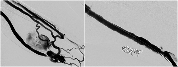
Dialysis is a process that filters blood in patients with kidney failure. This process is typically achieved through an arteriovenous fistula (or graft) created in the arm and then connected to a dialysis machine which draws the blood from the fistula. An arteriovenous fistula is a connection that is made by surgery between an artery and a vein usually in the arm. A graft is a fabric tube connected to an artery and a vein in the arm. Arteriovenous fistulas require brisk blood flow through them to be effective for dialysis. Fistulas and grafts may develop a focal narrowing within a neighboring vein or artery called a stenosis. Sometimes, fistulas and grafts become clotted which blocks the flow of blood, preventing their use in dialysis. When an arteriovenous fistula or graft becomes dysfunctional, an ultrasound may be performed to help identify the problem. A stenosis or blood clot may be treated by an Interventional Radiologist during a minimally invasive procedure called fistulography. The procedure is performed by inserting a catheter into the arteriovenous fistula after application of a local anesthetic (lidocaine). Contrast is injected through the catheter to visualize the fistula and diagnose the underlying problem. If the fistula is clotted, the catheter may be used to infuse clot dissolving drugs or deploy a small device that mechanically breaks up the clots. If a focal narrowing is seen on fistulography, this may be corrected by dilating the vessel with a small balloon (angioplasty). Once the procedure is complete, the catheters are removed, and a small suture is used to close the skin. Repaired fistulas and graft are generally able to be used immediately after the procedure.
Sedation: Local anesthesia (lidocaine) and moderate sedation (fentanyl and midazolam).
Procedure time: 30 minutes.