Ultrasound Constrast
Contrast Enhanced Ultrasound
65 year-old man with tumor recurrence from colorectal carcinoma metastasis in the right hepatic lobe 12 months following radiofrequency ablation in another institution:
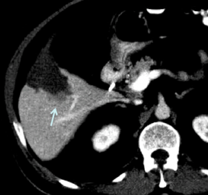
Axial contrast enhanced computed tomography demonstrates non-enhancing ablation zone in segment 5 with posteriorly adjacent tumor recurrence (arrow).

Positron emission tomography confirmed FDG-avid focus (arrow), corresponding to hypovascular metastasis on CT above.

B-mode (left) and color Doppler (right) ultrasound demonstrate tumor recurrence (arrows) poorly distinguishable from ablation zone.

Ultrasound contrast was used to guide the ablation needle confidently into the recurrent tumor portion next to the ablated area. Contrast enhanced ultrasound 22 seconds following injection of microbubbles (left) demonstrates clear contrast washout of vascular metastasis (arrows) adjacent to non-enhancing ablation zone in liver segment 5. Real time split screen view with B-mode image on the right is used to identify the area of interest before injection, which would otherwise be obscured by tissue cancellation techniques necessary to visualize microspheres in real time (see cine clip).

3 months following ultrasound-guided re-ablation of the recurrent tumor portion CT demonstrates successful result with complete ablation of tumor recurrence (left before, right after re-ablation).