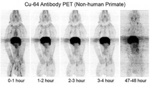
Translational Bioimaging Core
Mission
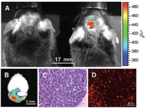
The Translational BioImaging Core Shared Resource (TBICSR) provides state of the art in vivo imaging equipment and expertise to support the diverse array of Consortium member preclinical research projects. TBICSR strives to provide easy access to a wide array of imaging modalities including magnetic resonance imaging (MRI), positron emission tomography (PET), computed tomography (CT), ultrasound (US), and optical imaging in a cost effective and efficient manner. Due to its multi-expert, multi-institutional organization, TBICSR bolsters the research objectives of Consortium Users through personnel with knowledge specific to each imaging modality and relevant in vivo biomarkers. TBICSR imaging modality experts, known as consultants in TBICSR, and imaging specialists direct Consortium users to the proper image modality through consultation and education. TBICSR staff foster animal imaging studies by providing scientific imaging and animal protocol development services, direct preclinical data acquisition, and image analysis and interpretation.
Services
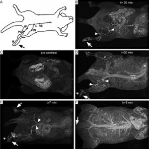
The TBIC supports and facilitates imaging research for members of the Cancer Consortium using resources available at Fred Hutch and UW in collaboration with multi-disciplinary imaging research groups specialized in respective imaging technologies. The TBIC provides services including:
- Support for animal imaging using equipment available to the Consortium
- Consultation for the effective use of imaging for specific research projects
- Training and education in imaging research
- Facilitation of the development of new imaging technologies
- Infrastructural support for animal imaging research
- Strategic acquisition of new imaging devices
Resources
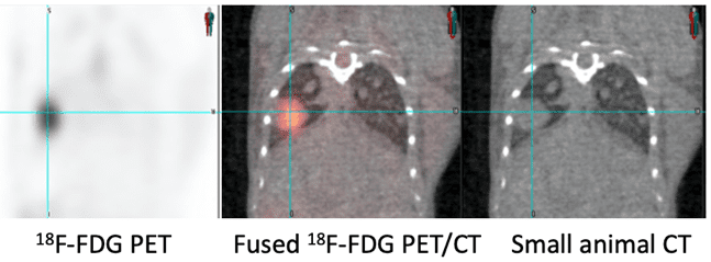
The TBIC Core resources are allocated at Fred Hutch. Other resources are accessible through centers, shared resources, and laboratories in the Department of Radiology, Washington National Primate Research Center, and the Department of Radiation Oncology.
|
Modality |
Equipment |
Location |
|
Optical |
IVIS Spectrum (x 2) |
Fred Hutch (FH) |
|
IVIS Spectrum* |
FH |
|
|
Lago-X* |
FH |
|
|
IVIS Spectrum (x2) |
UW South Lake Union (SLU) |
|
|
IVIS Spectrum* |
UW Animal Research and Care Facility (ARCF) |
|
|
Cryo-Fluorescence Tomography |
Xerra* |
UW ARCF |
|
Microscopy |
Two-photon Zeiss microscope + OPO |
FH |
|
Ultrasound |
Visual Sonics Vevo F2 ultrasound* |
FH |
|
Aixplorer/Verasonic software |
UW |
|
|
X-Ray |
MX-20 Specimen Radiograph |
FH |
|
Magnetic Resonance Imaging (MRI) and Magnetic Resonance Spectroscopy (MRS) |
Philips 3T Achieva (x 2) |
UW SLU and UW Medical Center |
|
7T/3T MRI MRS* |
FH |
|
|
4.7T Varian MRI/MRS |
UW |
|
|
14T MRI/MRS |
UW SLU |
|
|
Positron Emission Tomography (PET) |
Hamamatsu PET |
UW |
|
Positron Emission Tomography/Computed Tomography (PET/CT) |
Siemens Inveon PET/CT |
UW ARCF |
|
External Beam Radiation Therapy (EBRT) with CT |
xstral Small Animal Radiation Research Platform (SARRP) plus small animal Proton Beam |
UW Radiation Oncology |
|
Computed Tomography (CT) |
Quantum GX2 micro-CT* |
FH |
|
Body Composition Measurement |
EchoMRI* |
FH |
Core Members
Robert Miyaoka, PhD (Director, UW Radiology)
Elena Carlson, MS (Manager of Preclinical Imaging, Fred Hutch Cancer Research Center)
Donghoon Lee, PhD (UW Radiology)
Neal Paragas, PhD (TBIC Faculty Consultant, UW Radiology)
Mark Muzi, MS (TBIC Scientific Consultant, UW Radiology)
Contact
Please contact Teri Blevins (tblev@uw.edu) for general questions.
