Vascular Imaging Laboratory
 Co-Director: Mahmud Mossa-Basha, MD, UW, Radiology, Email: mmossab@uw.edu
Co-Director: Niranjan Balu, PhD, UW, Radiology, Email: ninja@uw.edu
Senior Advisor: Chun Yuan, PhD, University of Utah; Professor Emeritus, UW, Email: cyuan@uw.edu
Senior Advisor: Tom Hatsukami, MD, UW, Surgery, Email: tomhat@uw.edu
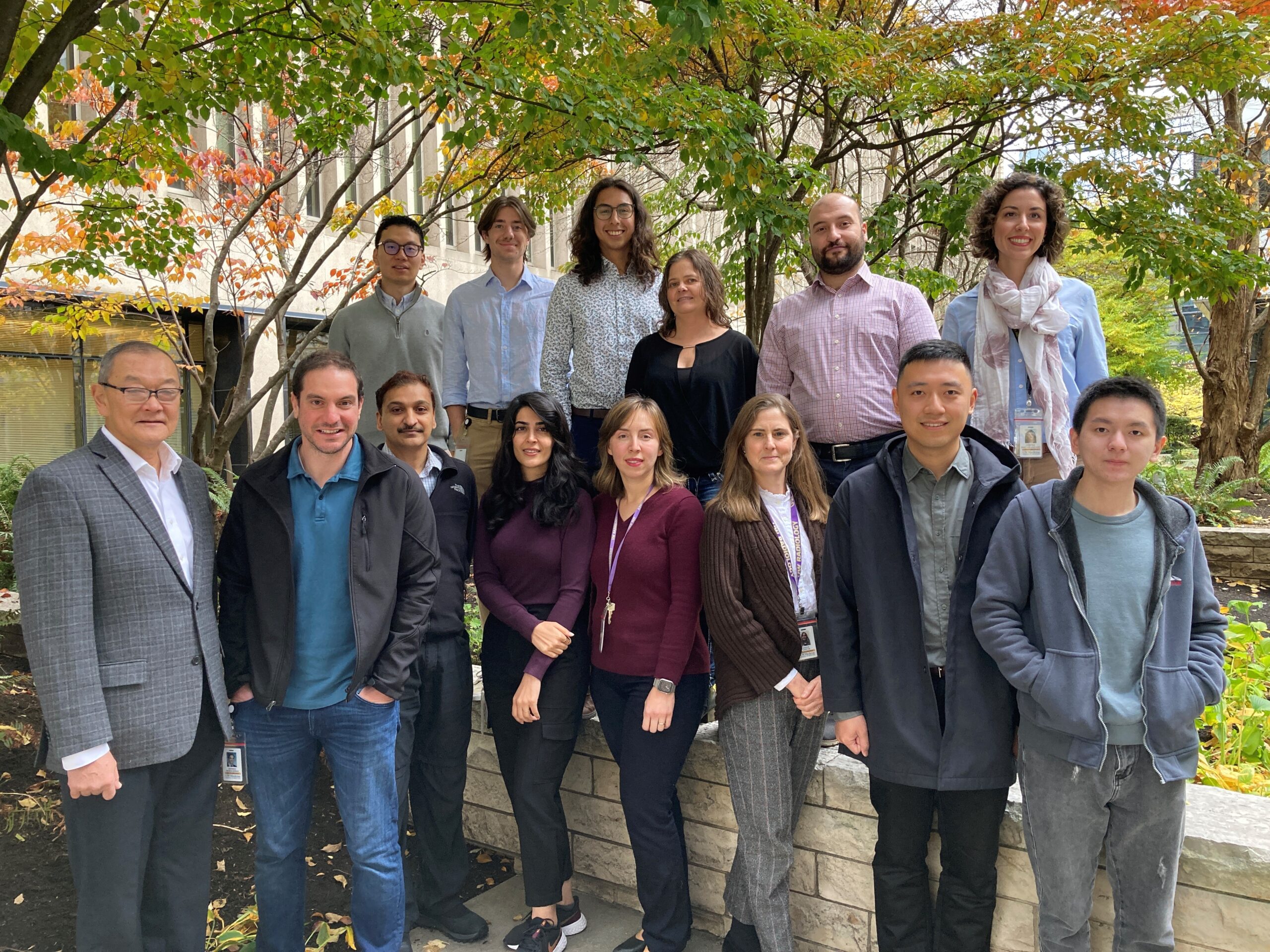
The Research Vascular Imaging Laboratory (VIL) at the University of Washington is comprised of a multi-disciplinary group of clinician scientists and researchers who are dedicated to advancing scientific knowledge, education, and training in the field of stroke and cerebrovascular disease, with a primary focus on atherosclerosis, intracranial vascular diseases, stroke and dementia research using high-resolution MRI and other imaging modalities such as CT, PET and ultrasound, while leveraging image processing and artificial intelligence. Our goal is to make advanced neurovascular imaging accessible and interpretable by all interested clinicians through improved post-processing and interpretation tools. We also provide training, pathology and clinical trial services.
Mission Statement
The Vascular Imaging Lab is committed to advancing imaging, post-processing and AI technology for identifying the underlying causes of cerebrovascular and cardiovascular disease initiation, progression, and complications.
VIL News
- In September 2022 the VIL held “The Foundations and Future of Vessel Wall Imaging” Symposium, successfully bringing together a large cohort of multidisciplinary speakers and participants, from Neurosurgery, Bioengineering, Electrical Engineering, Neurology and more. More details can be found here.
-
As the 2021 President Elect for the Society of Magnetic Resonance Angiography, VIL advisor and University of Utah Vice Chair for Research Dr. Chun Yuan and members of the VIL organized the successful international conference from September 10th-12th, 2021 with attendees from 21 countries.
A highlight of the conference was the Vessel Wall Segmentation Grand Challenge [Vessel Wall Segmentation Challenge (grand-challenge.org)] in partnership with MICCAI (Medical Image Computing and Computer Assisted Intervention) organized by Drs. Chun Yuan and Niranjan Balu from the Department of Radiology. The Grand Challenge aimed at automated segmentation of 3D carotid vessel wall MRI was successfully completed with the help of several members of the VIL. Over 200 teams from all over the world registered for the Challenge and 9 teams were invited to present their machine learning and deep learning based solutions at SMRA 2021 and MICCAI 2021. The winning team, WALL-E from the Department of Applied Mathematics at University of Twente, The Netherlands, received $4,000.
Drs. Chun Yuan and VIL Co-Director Mahmud Mossa-Basha from the Department of Radiology also mentored early career researchers in a fireside chat and delivered educational sessions talks on vessel wall imaging.
VIL also had a number of presentations at the SMRA, including:
Scientific Talks
Carotid Atherosclerosis is Associated with Brain Infarcts and Cognitive Decline – Niranjan Balu
Shorter Length of Visible Intracranial Arteries on Non-contrast Enhanced MRA is Associated with Cognitive Impairment – Zhensen Chen
Multi-planar, Multi-contrast and Multi-time Point Analysis Tool (MOCHA) for Intracranial Vessel Wall Imaging Analysis – Yin Guo
Educational Sessions
Clinical Implications of Vessel Wall Imaging and Applications – Mahmud Mossa-Basha
Intracranial and Extracranial Vessel Wall Imaging: How Do We Do It? – Baocheng Chu
Vessel Wall MR Imaging Techniques – Niranjan Balu
AI in Vascular Disease Screening and Large Clinical Trial Applications – Chun Yuan
- May 26th, 2021: Our work analyzing knee MR from the Osteoarthritis Initiative dataset has been featured in an Editorial in the Journal of the American Heart Association by Drs. David Bluemke and Nadine Kawel‐Boehm:
“Yuan et al used knee MRI examinations from the OAI in an unexpected manner—to design and test an AI to study the development of atherosclerosis of the popliteal artery. The characterization of peripheral artery disease (PAD) is a compelling topic equal to that of osteoarthritis. An estimated 27 million individuals in Europe and North America are affected by PAD, with more than 400 000 annual patient admissions attributed to PAD. Yuan et al evaluated more than half a million magnetic resonance MRI slices from 4688 study participants in the OAI. The extraordinary extent of data analysis was made possible by a deep learning algorithm that identified more than 9100 popliteal arteries (bilateral arteries were evaluable in most subjects). After finding the popliteal artery on the knee MRI, the algorithm preceded to determine three parameters: the popliteal artery lumen diameter, the wall thickness, and the total artery diameter.
An automated, AI‐driven analysis of biologically important arteries on this order of this magnitude has never been previously reported. The important implications of this work are wide‐ranging and multi‐factorial.“
The original JAHA manuscript can be read here: Atherosclerotic Burden and Remodeling Patterns of the Popliteal Artery as Detected in the Magnetic Resonance Imaging Osteoarthritis Initiative Data Set | Journal of the American Heart Association (ahajournals.org)
- Feb 1st, 2021: Dr. Chun Yuan and Dr. Xihai Zhao will jointly hold the 33rd Annual International Conference for The Society for Magnetic Resonance Angiography (SMRA) on September 10th – September 12th, 2021.
- Jan 15th, 2021: We are organizing the Carotid Artery Vessel Wall Segmentation Challenge supported by Society for Magnetic Resonance Angiography (SMRA). Welcome your participation.
- April 10th, 2020: Our book, Vessel Based Imaging Techniques, recently published by Springer, has received 4 stars from Doody’s Book reviews. “This book is of high quality and provides information that is essential to clinicians who use vessel-based imaging…I am not aware of a book that discusses imaging techniques based on different vascular beds the way this one does. Overall, this book does a great job in bridging the gap between research in current and new imaging techniques and clinical application.” Andres Guerra, MD (Northwestern University Feinberg School of Medicine)
- February 5th, 2020: Our team won the competition for artificial intelligence research by American Heart Association and Amazon Web Services through a collaborative data grant initiative (Automated Vessel Wall Screening to Predict Cardiovascular Risk) funded by AHA. Media coverage1. Media coverage2.
- October 28th, 2019: The Vascular Imaging Laboratory is proud to be working with the Department of Radiology on The Advanced Medical Imaging Program, an eight week summer exchange program for undergraduate students to experience research work. Applications will be opening in December 2019 for the summer 2020 program. Please see the link for more details. If you have questions please email Zach Miller zach1@uw.edu
- April 2019: Via an NIH R01 Grant, VIL and our collaborators have been developing new techniques for vessel imaging to look at vessels in the intracranial artery, to find associations between brain vessels and aging related diseases. The following papers have been recently published:
- VIL’s TOF MRA and iCafe technologies can provide a novel way to assess vascular condition and changes in intracranial vessels of older adults: Chen L, Sun J, Hippe DS, Balu N, Yuan Q, Yuan I, Zhao X, Li R, He L, Hatsukami TS, Hwang JN, Yuan C. Quantitative Assessment of the Intracranial Vasculature in an Older Adult Population using iCafe (intraCranial Artery Feature Extraction). Neurobiology of Aging. Available online 28 March 2019
- Contrast-enhanced IVW imaging, with the utilization of detailed plaque definition guidelines for image review, can be a reproducible technique for the evaluation of intracranial atherosclerosis: Mossa-Basha M, Watase H, Sun J, Shibata DK, Hippe DS, Balu N, Hatsukami T, Yuan C. Inter-rater and scan-rescan reproducibility of the detection of intracranial atherosclerosis on contrast-enhanced 3D vessel wall MRI. Br J Radiol. 2019 May;92(1097):20180973. doi: 10.1259/bjr.20180973. Epub 2019 Feb 26.
- CUSTOM + STEP allows multi-contrast 3D 0.5-mm isotropic Intracranial Vessel scans within 30 minutes: Balu N, Zhou Z, Hippe DS, Hatsukami T, Mossa-Basha M, Yuan C. Accelerated multi-contrast high isotropic resolution 3D intracranial vessel wall MRI using a tailored k-space undersampling and partially parallel reconstruction strategy. MAGMA. 2019 Jan 3.
- Intracranial artery features can be reliably quantified from TOF MRA using iCafe to provide both clinical diagnostic assistance and facilitate future investigative quantitative analyses: Chen L, Mossa-Basha M, Sun J, Hippe DS, Balu N, Yuan Q, Pimentel K, Hatsukami TS, Hwang JN, Yuan C. Quantification of morphometry and intensity features of intracranial arteries from 3D TOF MRA using the intracranial artery feature extraction (iCafe): A reproducibility study. Magn Reson Imaging. 2019 Apr;57:293-302.
-
Feb 28, 2019: The paper “Quantitative Assessment of the Intracranial Vasculature in an Older Adult Population using iCafe (intraCranial Artery Feature Extraction)” has been accepted in Neurobiology of Aging.
-
Jan 3, 2019: The paper “Accelerated multi-contrast high isotropic resolution 3D intracranial vessel wall MRI using a tailored k-space undersampling and partially parallel reconstruction strateg” has been accepted in Magnetic Resonance Materials in Physics, Biology and Medicine.
-
October 18th, 2017: Vascular Imaging Laboratory Member Dr. Dongxiang Xu was awarded the “Best Technology Innovation Award” from the 2017 Artificial Intelligence + Pharmaceutical and Healthcare Innovation Summit (AIPHIS), which took place on October 17th-18th in Shenzhen, China. The award was for the VIL technology package Vessel –AID, a comprehensive suite of software tools for vessel imaging analysis.
-
October 13th, 2017: VIL Co-Director Dr. Mossa-Basha and other VIL lab members Dr. Hatsukami, Dr. Balu. Daniel Hippe, and Dr. Yuan have received the cover image for their article “Added Value of Vessel Wall Magnetic Resonance Imaging for Differentiation of Nonocclusive Intracranial Vasculopathies” published in Stroke.
-
August 14, 2017: In collaboration with research colleagues at Tsinghua University, Tiantan Hospital, Beijing Hospital, Peking University First Hospital, Fujian Union Hospital, and the Hospital of Nanjing University Medical School, Vascular Imaging Laboratory Members Daniel Hippe, Dr. Gador M. Canton. Dr. Jie Sun, Dr. Kiyofumi Yamada, Dr. Dongxiang Xu, Dr. Thomas Hatsukami, and Dr. Chun Yuan have published the manuscript “Prevalence of Carotid Artery High Risk Atherosclerotic Plaque in Patients with Cerebrovascular Symptoms: A CARE-II Study” in the Journal of the American Heart Association.
- May 1st, 2017: Vascular Imaging Laboratory Symposium brings together international researchers. News
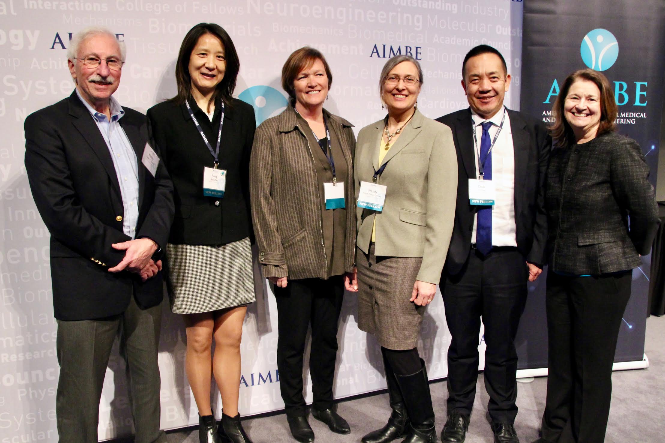
- March 20th, 2017: Dr. Chun Yuan has been inducted into The American Institute for Medical and Biological Engineering (AIMBE). (Photo)
- November 28th, 2016: Dr. Chun Yuan has received an Academy of Distinguished Investigators (DI) award.
- July 26th, 2016: Lab member Jin Liu has published a paper on Intraplaque Hemmorrhage detection in JCMR
- March 29, 2016: Journal of Magnetic Resonance Imaging (JMIR) selects VIL paper “Contrast-Enhanced High Resolution MRI for Atherosclerotic Carotid Artery Tissue Characterization ” as one of its 25 influential papers
- February 26th , 2016: Dr. Yuan nominated for AHA’s Clinical Research Prize for 2016.
- Feb 8, 2016: Lab member Kristi Pimentel has a five year work anniversary in our lab. Congratulations and thank you for your contributions, Kristi!
- Feb 1, 2016: Lab member Jin Liu’s abstract “Motion Detection and Correction for Carotid Artery Wall Imaging using Structured Light” is accepted by ISMRM 2016 as an oral presentation. Congratulations, Jin!
- Jan 21, 2016: Dr. Peter Börnert, Principle Scientist in Philips Research Hamburg, visits our lab for two days and gives a wonderful talk entitled “Selected MR-methodological activities at Philips Research Hamburg”.
- Jan 20, 2016: Lab member Dr. Jie Sun’s paper “Blood Pressure Is a Major Modifiable Risk Factor Implicated in Pathogenesis of Intraplaque Hemorrhage: An In Vivo Magnetic Resonance Imaging Study” is accepted by Arteriosclerosis, Thrombosis, and Vascular Biology. Congratulations, Jack!
- Jan 5, 2016: Dr. Xianling Wang from Xuanwu Hospital, China, joins the lab as a visiting scholar. Welcome, Xianling!
- Jan 5, 2016: Dr. Yuan presents at Imaging Sciences Research Lecture series on “Vessel Wall Imaging and International Studies”. RR-202 conference room at UWMC.
- Nov 30, 2015: Dr. Yao Shang from Beijing Tsinghua Chang Gung Hospital, China, joins the lab as a visiting scholar. Welcome, Yao.
- November 8, 2015: The Department of Radiology is proud to announce a new venture with the Center for Biomedical Imaging Research (CBIR) at Tsinghua University, a long-time collaborator with our lab, to explore formation of a joint research center to develop medical imaging technology. The center will involve multiple disciplines across different colleges, schools and hospitals and will bring together medicine, research and technology. (For more information, check this link.)
- Oct, 2015: We are proud to announce that our MRI system has been successfully upgraded to the state-of-the-art 3T Philips Ingenia MRI system. (Please check the details about the new scanner by this link.)