Metabolic Spectroscopy Laboratory
RESEARCH PROGRAMS IN THE METABOLIC SPECTROSCOPY LABORATORY
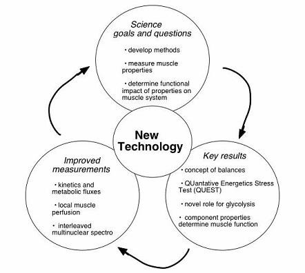
On this page brief descriptions of the three broad areas covered by our research programs are provided. More detailed information about these programs can be accessed by clicking on the tabs found on the left side panel.
ANIMAL RESEARCH
The animal research program uses three main approaches to study mitochondrial function in aging and disease:
- We use optical and NMR spectroscopy to measure mitochondrial function in vivo.
- We use genetically modified animals to manipulate in vivomitochondrial function.
- We combine systems analyses of in vivo energetics and gene expression data to study regulation of mitochondrial function.
HUMAN RESEARCH
 Our laboratory is a pre-eminent laboratory in developing state-of-the-art optical (OS) and magnetic resonance spectroscopic (MRS) methods for non-invasive measurements of metabolism and flow in human in vivo. Our work is focused on translating high-sensitivity spectroscopic tools and basic science insights of cell and vascular pathophysiology into new tools for disease diagnosis. This program in Metabolic Spectroscopy is complementary to Molecular Imaging approaches by uniquely providing high-sensitivity detection and in vivo quantitative measurement of key metabolic fluxes relevant to diabetes and metabolic disorders, cancer, aging and neurodegenerative diseases. Our work is directed to three goals.
Discovery – Identifying and quantifying key signaling molecules that regulate tissue function and underlie disease at the cellular level in vivo is made possible by high-sensitivity OS and MRS measurements. A current project is measurement of low concentration signaling molecules in brain centers regulating food intake, body weight and satiety. Identification of biomarkers in Huntington’s disease, ALS and aging is also in progress.
Development – New methods for direct measurement of cell metabolism, oxygenation and blood flow in vivo are developed by a multidisciplinary team composed of physicists, physiologists and physician-scientist. Recent advances include the first quantitative in vivo measurements of mitochondrial oxygen consumption and of tissue perfusion. A new research program focused on Mitochondrial Medicine has resulted from the insights gained from quantifying dynamic energetic fluxes and tissue blood flow. This work is focused on the role of mitochondrial dysfunction in aging, type 2 diabetes and neurodegenerative diseases in vivo in human tissues.
Diagnosis – An important shortfall in current clinical tools lies not only in technology by also our in understanding of cell pathophysiology. To overcome these limitations, we combine state-of-the-art non-invasive technology coupled with basic science studies of the physiology of disease to guide development of novel diagnostic tools. The tools are designed to 1) diagnose presymptomatic disease changes, 2) reveal the degree of disease progression, and 3) show the effectiveness of a intervention designed to treat the disease.
SPECTROSCOPY and IMAGING
A fundamental tenet underlying the Metabolic Spectroscopy Laboratory’s approach is that current gaps in clinical tools are the result of deficiencies in our current understanding of the basic science. Our research focuses on generating new insights from basic research in order to develop new spectroscopic methods and tools that may ultimately have clinical applications.
Magnetic resonance spectroscopy (MRS) and optical spectroscopy (OS) provide two of the most powerful tools for non-invasive investigation of tissue properties and metabolism. The potential application in clinical studies is vast but presently limited by lack of sufficient understanding of the basic cellular and tissue properties relevant to disease and by inadequate components of clinical MR instruments and FDA approved optical devices. Four primary avenues of investigation utilizing MRS have been established:
- Identifying the presence of specific metabolites in a tissue.
- Quantifying the content, and concentration, of specific metabolites.
- Measuring metabolic flux at a physiologically relevant time scale, seconds.
- Chemical shift imaging and spectroscopic imaging (CSI or SI) for “molecular imaging”