Teaching Files
Archived Case 46
Contributed by: Katherine E. Dee, MD -
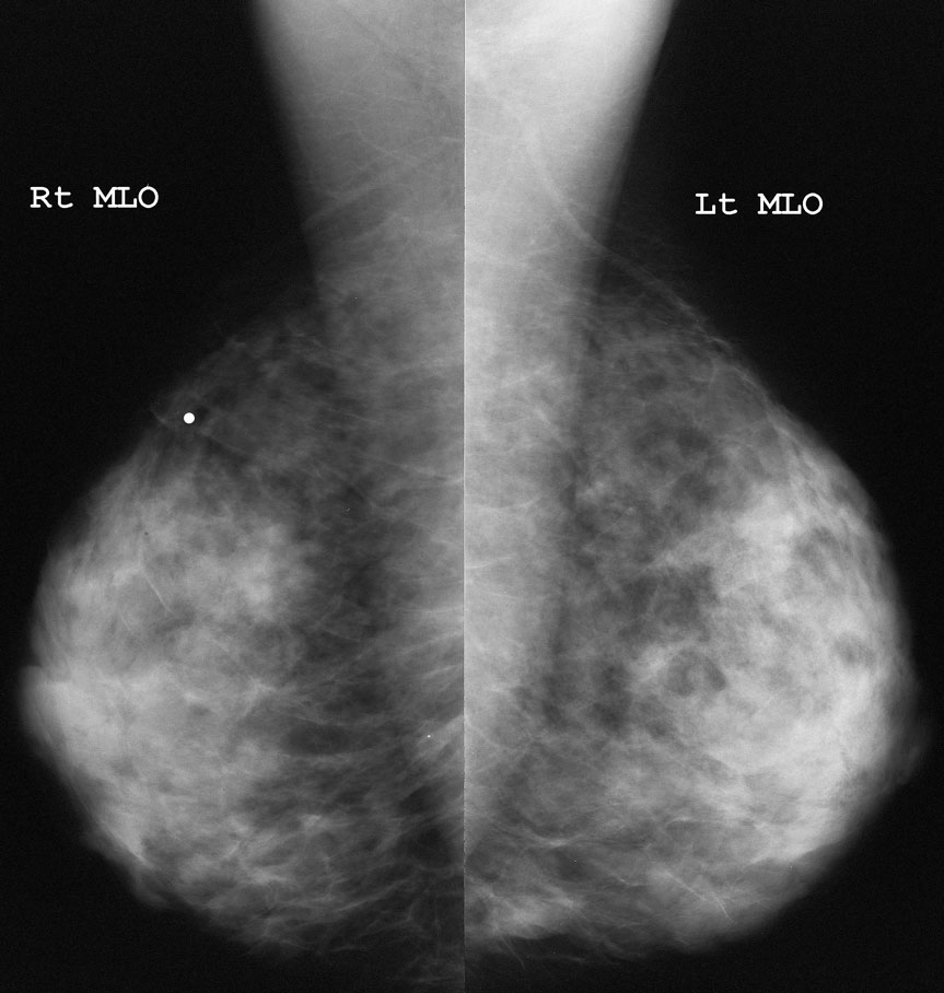
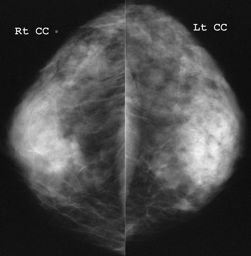
Palpable lump, right breast.
What would you do next?
Routine follow-up in 1 yearUltrasound
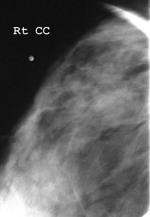
Spot views of the lump
MRI
Clinical follow-up for the lump
Now what do you recommend?
Routine follow-up in one yearUltrasound
MRI
Clinical follow-up of the lump
Biopsy
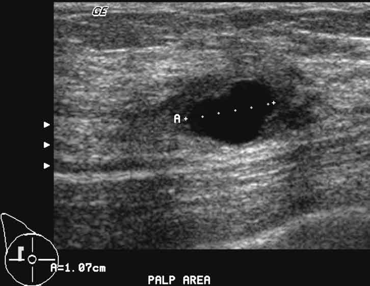
The tissue in this area is dense, and there is no definite abnormality seen on the spot compression views. Ultrasound is recommended for the work-up of any palpable abnormality unless the breast is entirely fat density in this location.
What is your assessment?
BI-RADS 1 - NormalBI-RADS 2 - Benign
BI-RADS 3 - Probably Benign
BI-RADS 4 - Suspicious
BI-RADS 5 - Highly Suggestive of Malignancy
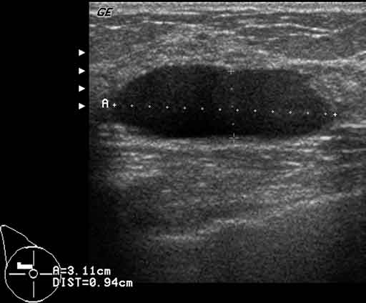
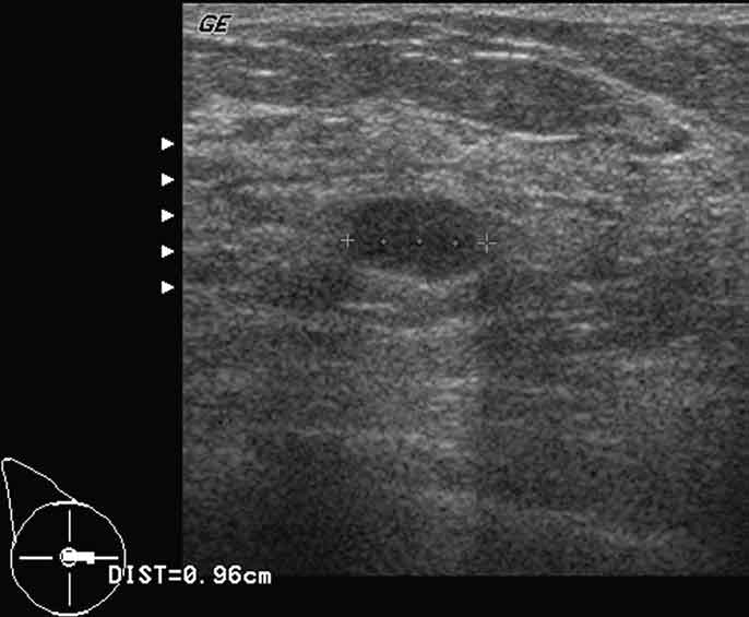
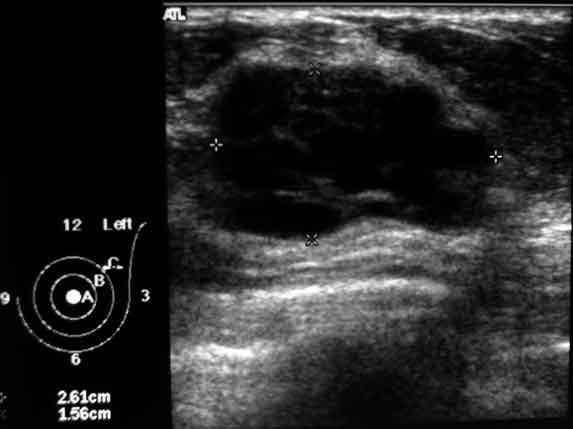
You are training a promising young breast imager, who says that this patient has multiple simple cysts in addition to the one described above, and shows you these images
What do you recommend now?
Return to routine screening mammography in 1 yearRecommend ultrasound in addition to screening mammogram in 1 year
Recommend aspiration of these additional cysts
Repeat the ultrasound yourself to confirm these cysts are simple
ultiple internal echoes make the diagnosis unclear. These could represent cysts or solid masses. The definition of a simple cyst requires the mass to be anechoic. Any jury can see from 30 feet that there are internal echoes in these masses.
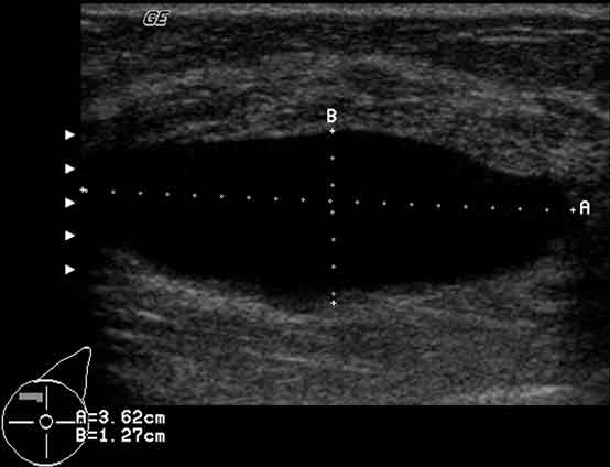
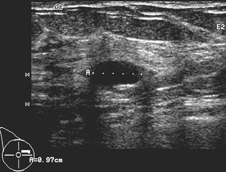
Repeat ultrasound shows
It is true that one can make anything anechoic if you turn down the gain enough. However, the gain can be turned down until the fat is appropriately hypoechoic. If the fat is anechoic, you’ve turned it down too far. Turning on harmonic imaging, as in the second image above, is often helpful to eliminate artifactual echoes.
What about this image?
What about this image?
BI-RADS 2 - Benign - Simple cyst
BI-RADS 3 - Probably Benign - Complicated cyst. Recommend 6 month follow-up ultrasound
BI-RADS 4 - Suspicious - Complex mass
recommend biopsy
