Teaching Files
Archived Case 10
Contributed by: Katherine E. Dee, MD -
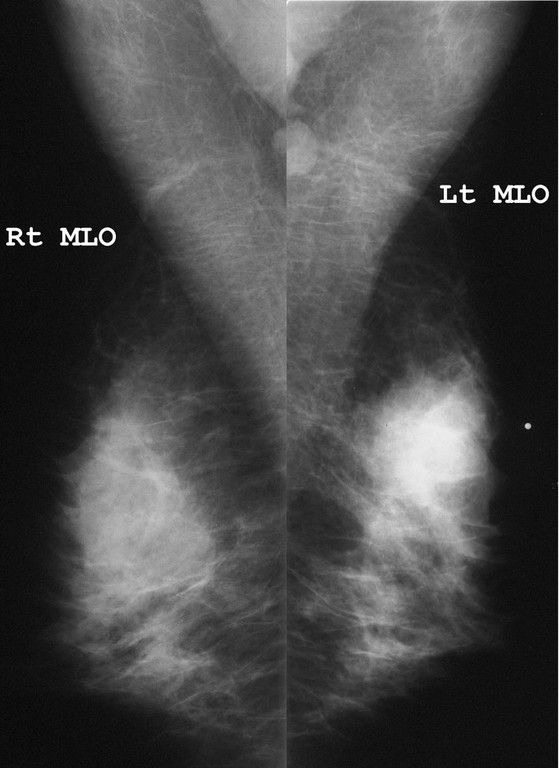
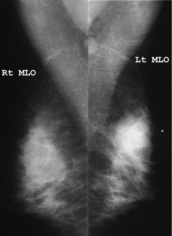
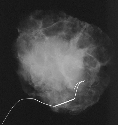
Would you perform an ultrasound?
Yes
No
A density is clearly present in the location of the lump. Unless the mammogram is totally fatty in this area, an ultrasound is indicated to evaluate the lump
What is your assessment?

Complex cyst
Mixed solid and cystic mass
Solid mass
There are multiple echoes at one end of the mass which were not artifactual. It is important not to overlook the solid-appearing portions of any cyst one is evaluating. [RSNA EJ 1999;3 Online.]
This patient underwent ultrasound-guided cyst aspiration which yielded yellowish fluid. Cytology was benign. The lump recurred soon after aspiration. Follow-up ultrasound examination showed the following:
What is the most likely diagnosis?
Cyst with hematoma from previous aspirationAbscess
Intracystic papillary carcinoma
Infiltrating ductal carcinoma with necrosis
Although the classic cystic cancer is Intracystic Papillary Carcinoma, they are very rare compared to the more common Infiltrating Ductal, NOS, and fluid aspirated from these tumors is usually bloody. This and the fact that this cystic mass previously yielded a negative aspirate make it more likely to be the more common IDC with necrosis. The necrotic center of a cancer is much more likely to yield a negative result. This is why core biopsy should be aimed at the solid portions of the mass.
Aspiration usually produces minimal bleeding, so a hematoma should be small and should be resolved by 3 months time. The patient had no symptoms of infection, and this nodular mass-like appearance would be unusual for an abscess.


