About Us
45 year old with chronic bilateral hearing loss for cochlear implant assessment
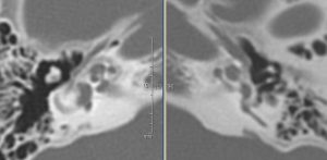
CT axial noncontrast bone windows
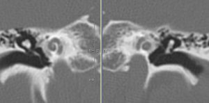
CT coronal reconstructions noncontrast bone windows
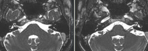
T2 axials
Findings:
CT: Subtle demineralization surrounding both the right and left cochlea, worse on the right side MRI: Increased T2 signal surrounding the cochlea bilaterally
DDX:
Cochlear otosclerosis
Diagnosis:
Cochlear otosclerosis
Discussion:
Cochlear otosclerosis is an uncommon cause of mixed and sensorineural hearing loss. It has a characteristic appearance on CT, producing a distinctive pericochlear hypodense double ring. However, its appearance on MRI is not as readily appreciated, producing a ring of intermediate signal in the pericochlear and perilabyrinthine regions on T2 and T1 weighted images sometimes with enhancement.
Submitted by Hari Challa, MD, UW Neuroradiology Fellow