About Us
74 y/o F progressive dysarthria, dysphagia.
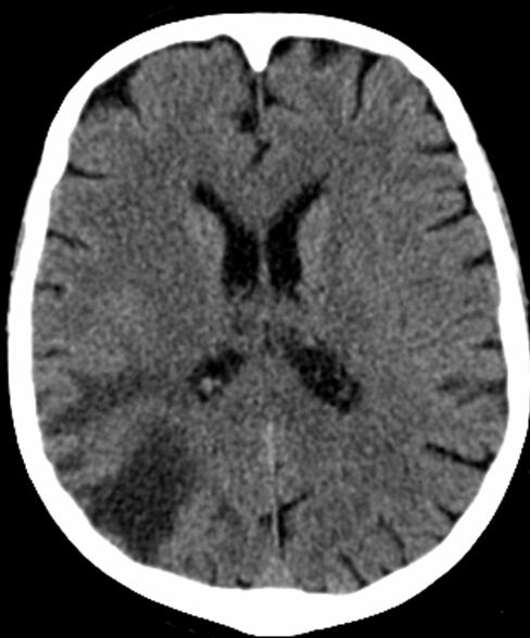
CT noncontrast 12/18/2008
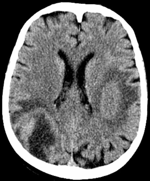
12/29/2008

Axial FLAIR 12/30/2008
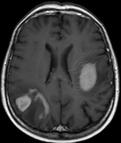
Axial T1 post Gad 12/30/2008
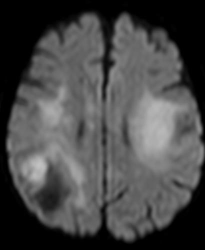
DWI

ADC map
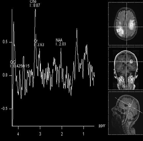
MRS TE=270
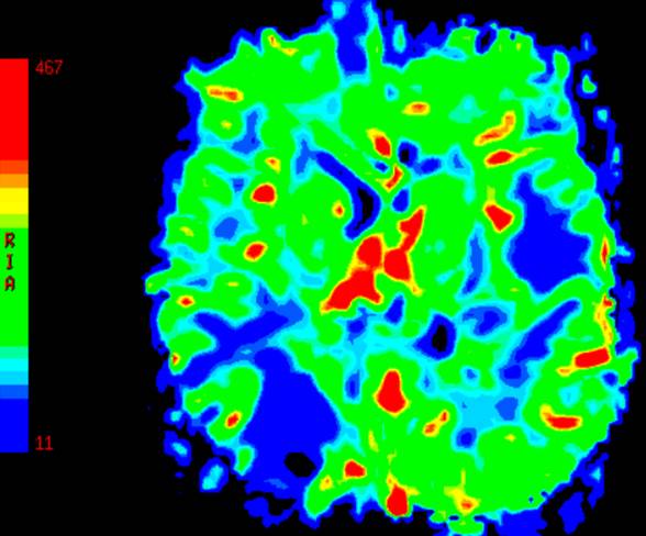
MR perfusion CBV map
Findings:
Multiple enhancing mass lesions with increased choline and restricted diffusion and minimally decreased perfusion
DDX:
Metastases, abscesses, lymphoma, demyelination
Diagnosis:
CSF analysis shows large cell B cell lymphoma.
Discussion:
Lymphoma with high cellularity may show restricted diffusion and iso or slightly decreased perfusion.
Submitted by Paritosh Khanna, MD, UW Neuroradiology Fellow