About Us
32 year-old with headache, fatigue, and neck pain and recent travel to India
FLAIR
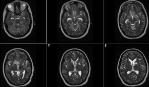
T2
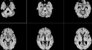
DWI
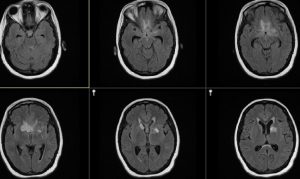
T1 noncontrast
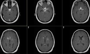
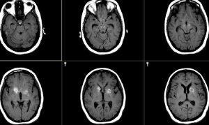
T1 post Gad
Findings:
Leptomeningeal enhancement filling the suprasellar cistern with extension up the perivascular spaces into the basal ganglia
DDX:
Granulomatous meningitis: Sarcoid, TB, fungus
Diagnosis:
TB
Discussion:
Infection begins in the lung, spreads to the bloodstream, then to the brain and meninges. Ventriculomegaly is present in 50-77% of patients. The basilar cisterns are filled with the purulent tuberculous exudate; marked enhancement is seen in this region following contrast administration. Infarcts (not seen in this case) can develop in the basal ganglia and thalami due to infiltration of the meningeal infection into the perivascular spaces resulting in vasculitis. The caudate nuclei, hypothalamus, and medial thalami are commonly affected. Punctate or ring-enhancing foci can be seen at the gray-white junction, less commonly dural based, which represent tuberculomas.
Submitted by Kathleen Tozer, MD, UW Neuroradiology Fellow