


UW Radiology contributes to international scientific statement on AI in cardiac imaging


UW Radiology Acting Instructor Domenico Mastrodicasa, MD, has published a scientific statement on artificial intelligence (AI) in cardiac CT and MRI in the journal Radiology. The statement is titled, "Use of AI in Cardiac CT and MRI: A Scientific Statement from the ESCR, EuSoMII, NASCI, SCCT, SCMR, SIIM, and RSNA."
In the scientific statement, Dr. Mastrodicasa and colleagues explored where AI is already making an impact in the cardiac imaging workflow, how ready these tools really are for clinical use, and what challenges still need to be addressed. As part of this effort, Dr. Mastrodicasa and colleagues adapted NASA’s Technology Readiness Level (TRL) scale, originally developed for space technology, to assess AI maturity in cardiac imaging. This scale looks like:
- TRL 1–2: Research phase (exciting ideas, but early days)
- TRL 3–6: Development (AI tools being tested and refined)
- TRL 7–9: Clinical deployment (ready, or almost ready, for real-world use)
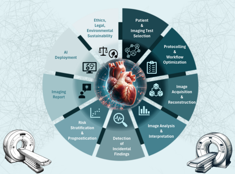
Figure 1
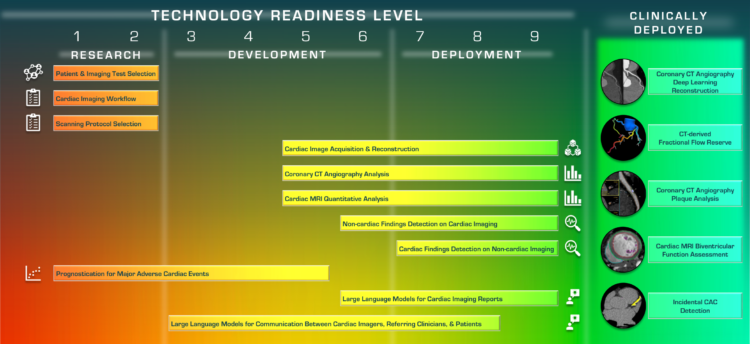
Figure 3
By evaluating AI through this scale, this work bridges the gap between innovation and clinical implementation. Beyond assessing AI readiness, the statement takes a holistic approach, addressing key considerations such as ethics, fairness, transparency, bias mitigation, and sustainability. Key takeaways and recommendations grounded in the collective expertise of the team of authors are summarized in Figure 4.
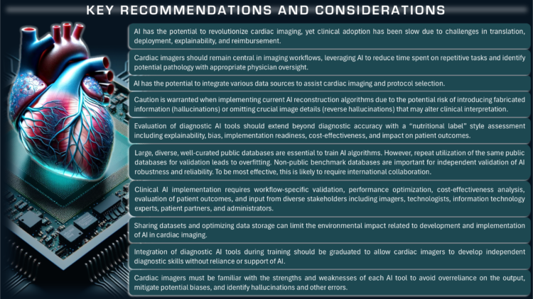
Figure 4.
The statement generated strong interest, with over 2,300 downloads in the first two weeks and an Altmetric score of 60, ranking among the top 5% of all research outputs tracked by Altmetric. Radiology Editor-in-Chief Linda Moy praised the article, recognizing its contribution to the growing conversation on AI in cardiac imaging. Engagement has also been particularly strong on LinkedIn, where the post has reached nearly 6,000 members and generated over 9,500 impressions. Following its publication, the statement has been featured in two AuntMinnie.com articles: RSNA and collaborators release statement on AI for cardiac CT, MRI and International collaboration leads to statement on AI for cardiac CT, MRI.
Dr. Mastrodicasa and co-authors hope this statement serves as a guiding framework to help cardiac imagers, AI experts, and policymakers work together to advance AI in clinical practice responsibly and effectively.
This work is the result of a multi-institutional collaboration among experts from leading institutions, including Marly van Assen, PhD (co-first author), from Emory University, Atlanta, Georgia; Merel Huisman, MD, PhD, from Radboud University Medical Center, the Netherlands; Tim Leiner MD, PhD, and Eric Williamson MD, from Mayo Clinic, Rochester, Minnesota; Edward D. Nicol MD, MBA, from the Royal Brompton Hospital, London, United Kingdom; Bradley Allen, MD, MS, from Northwestern University Feinberg School of Medicine, Chicago, Illinois; Luca Saba, MD, from University of Cagliari, Cagliari, Italy; Rozemarijn Vliegenthart, MD, PhD, (co-senior author) from University Medical Center Groningen, the Netherlands; and Kate Hanneman, MD, MPH, from University of Toronto, Ontario, Canada.
Listen to Dr. Mastrodicasa discuss this work in a podcast produced by Radiology entitled Radiology & AI: The Future of Cardiac Care.