Dr. Mona Kharaji recounts presenting on vessel wall imaging at RSNA 2024
By Mona Kharaji, MD, Neuroradiology Research Fellow
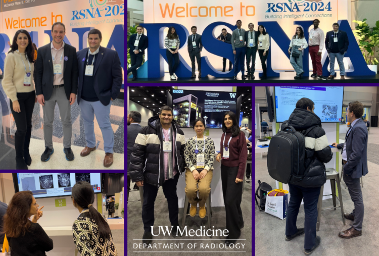
 Attending the RSNA 2024 Annual Meeting in Chicago was an incredible and enriching experience. Thanks to the generous support of the Gamble Cerebrovascular Research Fund and the Education Endowment Committee, I had the opportunity to present two educational exhibits focused on vessel wall imaging in stroke workup and embolic stroke of undetermined source, alongside my teammates: Dr. Javid Azadbakht, Dr. Ahmed Safwat and Dr. Maoxue Wang at the Vascular Imaging Lab (VIL).
Our exhibits, “Optimizing Vessel Wall Imaging for Cerebrovascular Assessment and Integrating it into Stroke Workup Standards of Care” and “Identifying and Characterizing Non-stenotic Potential Arteriogenic Sources of Emboli in Patients with Embolic Stroke of Undetermined Source,” showcased innovative techniques to improve stroke diagnosis. Representing our team’s collaborative efforts to a global radiology audience was an invaluable experience.
Beyond the opportunity to share our work, attending the conference allowed me to explore the latest advancements in radiology, from cutting-edge imaging methods to the integration of AI in clinical practice. Engaging with global experts and peers provided valuable insights and inspiration for future research projects.
I am sincerely grateful for the support that made this experience possible.