UW Radiology
Case 1
65 yr old female with liver mass suspected to be a HCC, no history of hepatitis or cirrhosis. The liver mass was diagnosed after a non contrast CT was performed for a minor car accident. CT with contrast was performed. Images given below.
| Arterial phase | Portal venous | Delayed phase |

Note the peripheral mild enhancement and irregular margins of the lesion. |

Portal venous phase – increasing peripheral enhancement |

Delayed phase |

Second lesion in the left lobe |

Portal venous phase |

Delayed phase |
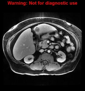
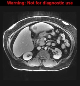
MRI of the abdomen:
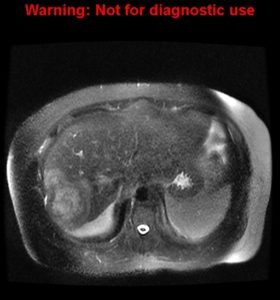
T2 weighted image |
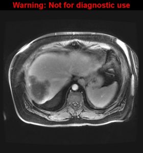
T1 pre contrast |
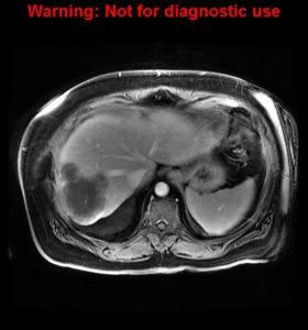
Arterial phase |
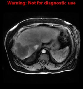
Venous phase |
2nd lesion |
Delayed phase |