Muscle Atlas
Tibialis Posterior
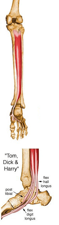
Origin: Posterior aspect of interosseous membrane, superior 2/3 of medial posterior surface of fibula, superior aspect of posterior surface of tibia, and from intermuscular septum between muscles of posterior compartment and deep transverse septum
Insertion: Splits into two slips after passing inferior to plantar calcaneonavicular ligament; superficial slip inserts on the tuberosity of the navicular bone and sometimes medial cuneiform; deeper slip divides again into slips inserting on plantar surfaces of metatarsals 2 - 4 and second cuneiform
Action: Principal invertor of foot; also adducts foot, plantar flexes ankle, and helps to supinate the foot
Innervation: Tibial nerve (L4, L5)
Arterial Supply: Muscular branches of sural, peroneal and posterior tibial arteries
The medical illustrations contained in this online atlas are copyrighted © 1997 by the University of Washington. They may not be utilized, reproduced, stored, or transmitted in any form or by any means, electronic or mechanical, or by any information storage or retrieval system, without permission in writing from the University of Washington.
Receiving a license to use these images is generally quite easy, particularly for academic and scholarly purposes. For more information see the Musculoskeletal Atlas Express Licensing Page.