About Us
77 year-old with recent syncopal episode
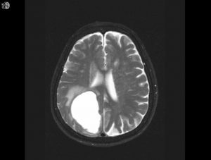
Axial T2
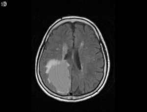
FLAIR
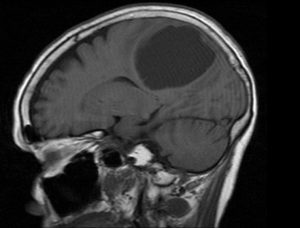
T1 sagittal noncontrast
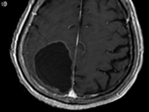
axial postcontrast
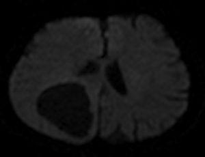
DWI
Findings:
Hyperdense mass with hyperostosis on NECT Homogeneously enhancing extra-axial dural based mass Cortical buckling and trapped cortical vessels
DDX:
Cystic brain tumors: pleomorphic xanthoastrocytoma (PXA), pilocytic astrocytoma, hemangioblastoma, ganglioglioma/gangliocytoma Metastases (particularly squamous cell and adenocarcinoma) Abscess particularly echinococcus if thin wall
Diagnosis:
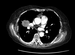
Metastasis from squamous cell carcinoma of lung
Discussion:
Although an abscess or primary tumor could not be excluded, the thin rim of enhancement and patient age with moderate surrounding edema favor a cystic metastasis.
Submitted by John Rhee, MD, UW Neuroradiology Fellow