About Us
42 year-old with headache
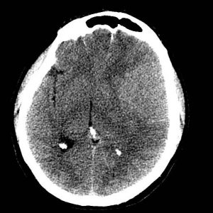
CT noncontrast soft tissue window
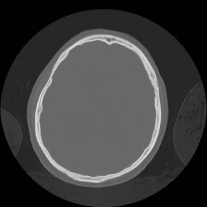
CT noncontrast bone window
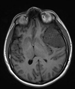
T1 axial noncontrast
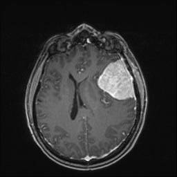
T1 axial post gad
Findings:
Hyperdense mass with hyperostosis on NECT
Homogeneously enhancing extra-axial dural based mass
Cortical buckling and trapped cortical vessels
DDX:
Dural metastasis
Granuloma (Sarcoid, TB)
Hemangiopericytoma
Solitary fibrous tumor
Diagnosis:
Meningioma
Discussion:
Arise from arachnoid meningothelial “cap” cells
Most common primary adult intracranial tumor
F>M; may be related to female sex hormones
XRT pre-disposes
Associated with NF-2
Dural based mass with cortical buckling and/or trapped CSF clefts/cortical vessels
Hyperostosis of bone; Calcifications
MRS: elevated levels of alanine at short TE
Submitted by Sung Logerfo, MD, UW Neuroradiology