Teaching Files
Case 3
Contributed by: Steven J. Rockoff, MD and Diana L. Lam, MD - June 1, 2020
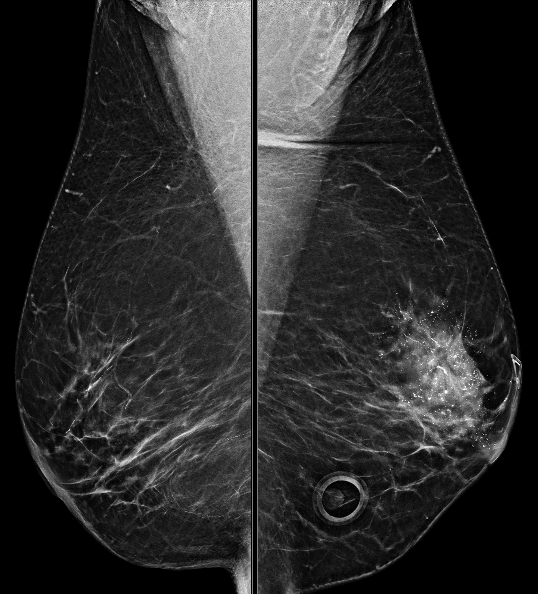
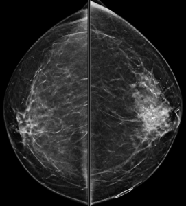
A bilateral diagnostic mammogram is performed for a 56-year-old woman with a left breast palpable abnormality and pain:


Other than these whole breast views, what additional views would you like to see in order to make your assessment of the dominant abnormality?
A. Tangential viewsB. Spot Magnification views
C. Cleavage views
D. Eklund views
Explanation: There are highly suspicious calcifications in the left breast. In order to best assess the calcifications, spot magnification views are needed, which will allow for a better description of the calcifications’ morphology and distribution.
Tangential views are used to determine whether a mammographic finding is located in the skin, such as skin calcifications.
Cleavage views are used to view the more medial breast tissue of both breasts.
Eklund views, also known as implant-displaced views, are obtained in order to view more breast tissue in women who have breast implants.
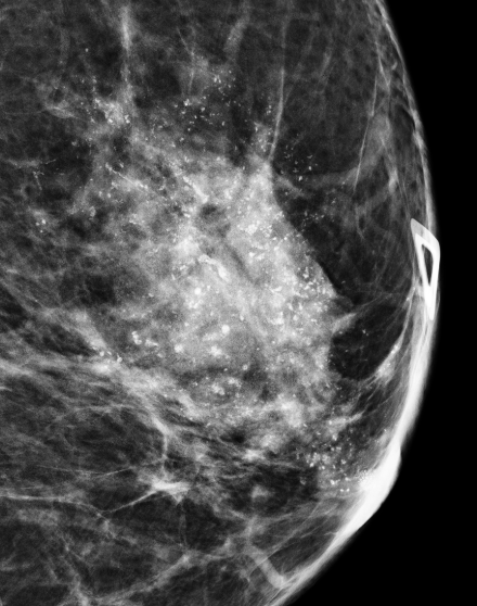
A spot magnification view in the ML projection is obtained:

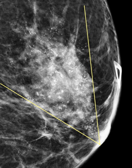
How do you describe the distribution and morphology of the calcifications?
A. Grouped distribution; Coarse heterogeneous morphologyB. Grouped distribution; Fine pleomorphic morphology
C. Segmental distribution; Coarse heterogeneous morphology
D. Diffuse distribution; Amorphous morphology
Explanation: These calcifications are in a segmental distribution, which is a roughly triangular shape which points towards the nipple. This pattern suggests that the calcifications are primarily located within the breast’s ductal system. The individual calcifications predominantly have discrete irregular shapes, are slightly coalescent, and are slightly larger than the fine pleomorphic type of calcification.

What is your assessment and recommendation?
A. BI-RADS 0 (Incomplete); Diagnostic USB. BI-RADS 2 (Benign); One year follow-up
C. BI-RADS 3 (Probably Benign); Six month follow-up
D. BI-RADS 4 (Suspicious); Stereotactic biopsy
E. BI-RADS 5 (Highly Suggestive of Malignancy); Stereotactic biopsy
Explanation: Either BI-RADS 4 or BI-RADS 5 could be an acceptable assessment for this patient. Stereotactic biopsy is usually the best way to obtain reliable tissue sampling in the setting of suspicious calcifications. You may have noticed the large focal asymmetry which is associated with the calcifications; in such a case, diagnostic ultrasound to look for a target that could be biopsied with ultrasound guidance would also be a reasonable option.
You decided to give the left breast BI-RADS 5 in your report. Stereotactic biopsy was performed. There are a large amount of calcifications in the biopsy specimen. The pathology report states that there are fibrocystic changes and other benign findings. What would you say for radiology-pathology concordance in the addendum to the biopsy report?
A. Discordant biopsy; Recommend surgical excisionB. Discordant biopsy; Recommend six month follow-up
C. Concordant biopsy; Recommend surgical excision
D. Concordant biopsy; Recommend six month follow-up
Explanation:
Segmental distribution has the highest likelihood of malignancy out of all of the calcification distribution descriptors, and coarse heterogeneous morphology is also considered suspicious. Coupled with the fact that there is a focal asymmetry associated with the calcifications, BI-RADS 5 is appropriate. If a BI-RADS 5 (Highly Suggestive of Malignancy) assessment is made, you must be prepared to recommend re-biopsy or surgical excision if the pathology report of the initial biopsy only describes benign findings.
A concordant biopsy means that the pathology findings are a reasonable explanation for the imaging findings. A discordant biopsy (as in this situation) means that the benign pathology findings do not adequately explain the suspicious imaging findings.
In reality, biopsy of this patient yielded invasive ductal carcinoma with ductal carcinoma in-situ (DCIS).