MRI Coil Laboratory
About the Lab
Dr. Cecil Hayes, a pioneer in RF coil physics, founded the RF Coil Laboratory in 1992. With the assistance of engineers Mark Mathis and Tim Wilbur the laboratory has produced numerous custom RF coils for research and clinical projects at the UW and other off-campus sites.
These include coils for high resolution imaging at 1.5T and 3T of the temporal lobes of the brain, the brachial plexus, peripheral nerves, the wrist, the carotid arteries, the neck, the torso, the pelvis, the heart, and for fMRI brain studies.
Special coils have also been produced for our research affiliates in Seattle; Children’s and Puget Sound Veterans Administrationhospitals. Experimental coils supporting high field, 4.7T and 7T scanners at the UW are also designed and supported, as well as custom coils for animal imaging at various field strengths.
In addition to RF coil design and construction, the laboratory scientists also develop mechanical and electronic accessories that are utilized for specific experiments in the high magnet field environment of the MRI scanners. RF Coil Lab scientists also design, build, modify, and maintain the electronic hardware for
the MR Research Laboratory’s 4.7T and 7T MR systems.
Several inventions, patents and applications, and commercial interests have been realized as a result of Coil Lab activities. All coils for human studies conform to US Food and Drug Administration (FDA) and Institutional Review Board (IRB) guidelines. For more information about our coils, please contact the DISC lab.
What is an RF Coil?
The quality of an MR image and the accuracy of the quantitative information it contains is limited by the degree to which the desired signal intensity exceeds the random noise that is also present. A radio frequency (RF) receiver coil picks up both the MR signal, which is a weak oscillating magnet field from the rotating hydrogen nuclei in the patient, and the noise, which is due to the random motion of charged particles in the imaging system. The RF receiver coil is basically a sophisticated antenna that functions as the imaging detector and is the critical element that conveys imaging information from the patient to the scanner. As such, its performance largely determines the signal-to-noise ratio (SNR) in the resulting image. High SNR can be used to reduce the time needed to acquire an image, to see smaller features of the anatomy (improve resolution), or to distinguish subtle variations in image contrast due to disease. The focus of the RF Coil Laboratory is to design RF coils that maximize signal and minimize noise picked up when imaging a subject. Often this improved SNR is the key requirement for making a new diagnostic or research procedure possible.
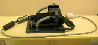
Mechanical devices associated with RF Coils that provide comfort and restrict patient movement during scanning are of particular interest because patient 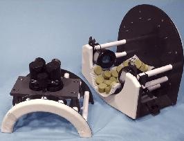 motion is a major cause of reduced image quality. For functional MRI studies of brain activity, sound and video devices have been built and then integrated into custom high-resolution head coils. The RF lab also provides electronic and mechanical design support for the UW MR Research Laboratory and has built devices for physiologic monitoring, stimulus response, and temperature control in the magnetic environment.
Can you build us a coil?
The Coil Lab has the ability to customize a variety of different coils and we pride ourselves on our ability to meet the needs of MR researchers and are dedicated to the challenges of modality customization. Feel free to contact the DISC lab and we’d be happy to discuss your needs.
Custom Coils Available
- Finger coils
- Mouse coils
- Rat brain and body coils
- Primate coils/Flex coil for primate heads
- 8 coil Neonatal Primate
- 8 Channel Carotid coil
- Neurovascular Carotid Coil