About Us
37 year-old with intractable epilepsy and inconclusive EEG
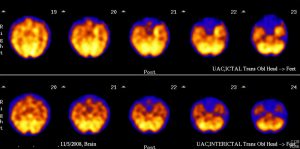
HMPAO SPECT axial images: Ictal (top) and interictal (bottom)
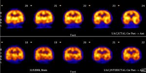
HMPAO SPECT coronal images: Ictal (top) and interictal (bottom)
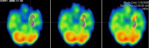
HMPAO SPECT axial images: Ictal vs Interictal subtraction images
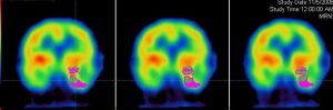
HMPAO SPECT coronal images: Ictal vs Interictal subtraction images
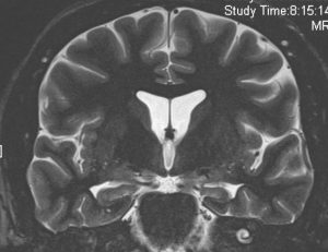
Coronal T2
Findings:
SPECT: Hyperperfusion on ictal SPECT in the left hippocampus MRI: Left hippocampal atrophy
DDX:
Left mesial temporal sclerosis
Diagnosis:
Left mesial temporal sclerosis
Discussion:
Many theories for the cause of mesial temporal sclerosis have been proposed but it is most likely related to uncontrolled febrile convulsions in childhood. Interictal and ictal brain SPECT can be performed with Tc99m labeled HMPAO or ECD. These both approximate regional cerebral blood flow. Brain SPECT performed using both interictal and ictal data as well as software to compare them directly is over 90% sensitive for temporal lobe epilepsy. The indications are when the seizure protocol MR is normal, the EEG results are inconclusive or discordant with the MR. On MRI coronal T2 and STIR thin section images increased signal, volume loss, loss of internal morphology are the typical finding in hippocampal sclerosis.
Submitted by Jennifer Kohr, MD, UW Radiology Resident